Health & Wellbeing
Our experts show you simple and effective ways to stay healthy and active, so you can live your passions every day.

Sunstroke and heatstroke: symptoms, signs and treatment
Sunstroke and heatstroke can be life-threatening - and we're at increased risk as we get older.

How to sleep well in the heat at home and on holiday
Expert advice on how to get a good night’s sleep when it’s hot – and 3 mistakes to avoid.

Why do my ankles swell in hot weather?

Exercising in the heat: How to stay safe
When hot weather hits it can be a challenge to exercise in the heat. We've got advice from the experts to help you keep active safely.

Should you brush your teeth before or after breakfast?
Dr Mark Porter settles a family debate of when we should brush our teeth in the mornings.

Why you feel healthier on holiday
The simple habits you pick up on holiday can make a real difference back home.

5 signs you could be stuck in a rut
If every day feels the same these expert tips can help you break the cycle.
Welcome to Experience is Everything – The Health Edition
Dr Mark Porter dives into the health issues affecting us all in the new podcast from Saga.
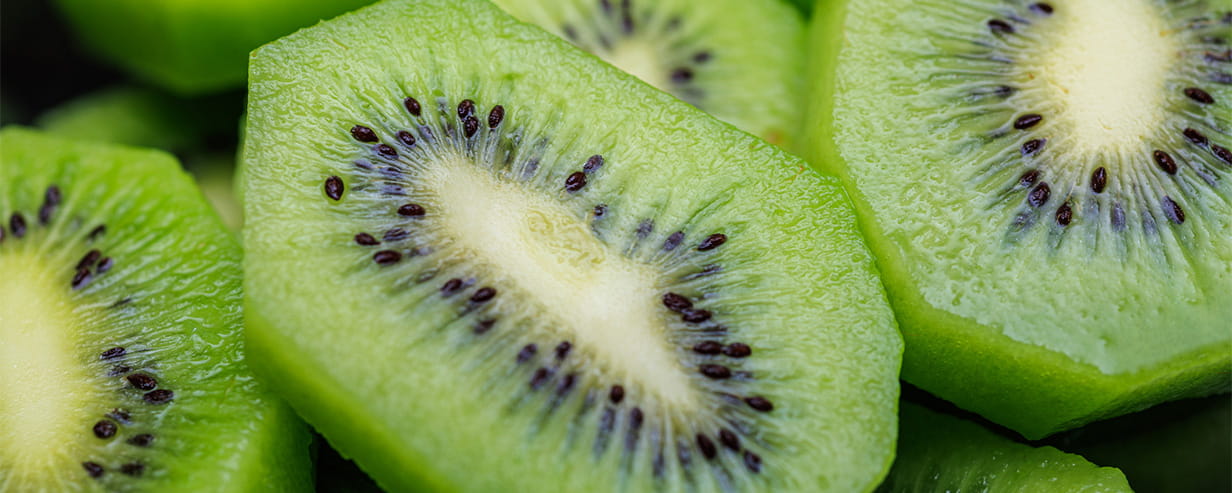
Why kiwifruit could be one of the healthiest fruits you can eat
Better sleep, digestion and moods can be some of the health benefits of kiwifruit.
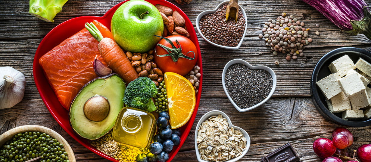
The 6 everyday foods that lower your risk of a heart attack
The simple dietary tweaks that experts say will cut your chance of cardiac arrest.

Mistakes you make talking to the doctor
Here’s how to get the most out of your appointment – and talk so your doctor will listen.

Are you making these common sunscreen mistakes?
From forgetting your ears to thinking one application lasts all day, these common SPF mistakes are putting older skin at risk.

Which supplements for joint pain actually work?
They’re all touted as cures for achy joints but which ones do the experts back?

How to create a routine in retirement
Our expert outlines the best way to bring structure, purpose and freedom to your days.

This year, Saga is celebrating an incredible 75 years – and we’d love you to be part of the celebrations. For a limited time only, you can subscribe to Saga Magazine for just 75p an issue.
Receive the next 6 print editions delivered direct to your door, plus 6 months’ unlimited access to the Saga Magazine app – perfect for reading on the go.
Play our free daily puzzles
Beat the boredom and exercise your mind with our selection of free puzzles.